Archive for the “Virology” Category
 Aethlon Medical has developed a new device called Hemopurifier® which acts a lot like the usual hemodialysis machine used in patients with end-stage renal disease but targets a different kind of particle within the blood, it captures viruses! The machine uses thin filters to capture & remove viruses from the blood. This requires an artery to act as an entry point of the blood to the machine, where it is filtered, and then sent back to the body, only cleaner. Aethlon Medical has developed a new device called Hemopurifier® which acts a lot like the usual hemodialysis machine used in patients with end-stage renal disease but targets a different kind of particle within the blood, it captures viruses! The machine uses thin filters to capture & remove viruses from the blood. This requires an artery to act as an entry point of the blood to the machine, where it is filtered, and then sent back to the body, only cleaner.
The whole blood circulation passes through the machine almost once every 8 minutes. The entire process itself requires a few hours. Needless to say, this might prove to be revolutionary in the treatment of all sorts of viral infections: measles, mumps, hepatitis, west-nile virus, smallpox, HIV, avian flu, even the seemingly harmless human flu..just to name a few.
The device has already received a lot of attention & was in fact awarded. In a pre-clinical study, an astonishing 99.4% of H5N1 flu virus, as verified by real-time PCR, was eliminated from the patient’s blood within an operating time of 6 hours.
This state-of-the-art device functions through using antibodies to capture viruses & toxins before their actual attack on the human organs. Patients can even be started on the machine before the physicians find out the cause of the disease.
Source: ScienceDaily
Tags: blood, H5N1, hemopurifier, HIV, immunotherapy, virus
 10 Comments »
You may remember HBV, the famous hepatitis virus with its partially double-stranded circular DNA genome. I always wondered: What is that supposed to mean?! HBV has a very complicated replication cycle. I’m pretty sure that all molecular biology fans will be totally thrilled by reading this.
HBV replication cycle is divided into 3 stages:
1- The infectious virion containing the partially double-stranded circular DNA, they call it RC-DNA (relaxed circular).
2- Right after the infection, inside the host nucleus, the genome becomes cccDNA (covalently closed circular DNA). It looks just like plasmids. HBV needs that highly stable form because it’s a chronic infection; it doesn’t want to be lost during host cell division. It may be still there in the host cells even after effective antiviral therapy.
3- Finally transcription takes place, several RNA molecules are produced, some of them are genomic (contain the whole genome) named pgRNA (pregenomic RNA) & some are subgenomic (encode needed enzymes) It uses the cell’s RNA polymerase II to do all this.

So, what happens to the pgRNA? They get inside progeny capsids ready to be reverse transcribed with the help of P protein (Its reverse transcriptase) which is “co-packed” in the pgRNA- progeny capsid package to get it back to the RC-DNA. Then the mature RC-DNA containing-nucleocapsids could undergo cccDNA amplification, or could be enveloped & ready for release from the cell. Of course all this is in equilibrium; if there’s only one copy in the cell, the priority is not to make cccDNA but to be enveloped & released.
Why the RC-DNA needs to be first cccDNA before transcription? As I got from this review, the RC-DNA has the normal (-)-strand (opposite sense to mRNA) but its complementary, the (+)-DNA strand, is not in full length. It results from the non-identical nucleotides supply; because the envelop is impermeable to nucleotides. At the 5′ end of the (-)-strand, there’s the P protein. But at the 5′ end of the (+)-strand, there’s some RNA nucleotides remains from the pgRNA…It was its primer, remember? All these are removed to be a cccDNA. The P protein may has a role in completing the (+)-strand.
Image credits:
Hepatitis B Virus Replication: http://www.meds.com/
Tags: cccDNA, HBV, P protein, pgRNA, RC-DNA, reverse transcription
No Comments »
Scientists are harvesting all of them potatoes for an investigational experiment is being done on patients with Alzheimer’s disease using protein extracts obtained from a potato virus.
Alzheimer’s is associated mainly with amyloid plaque within the neurons of the brain. A major portion is formed of beta amyloid which should, in normal cases, break down on its own but rather tends to accumulate forming the insoluble hard plaque. Here is where the potatoes pitch in.
A fairly known potato virus “PVY“, basically harmless to humans, which I & probably you might have been previously exposed to, contains an amyloid-like protein. Through isolating the potato virus & injecting it in experimental animals with booster doses every month, the levels of antibodies against the protein, in 4 months, quickly rose to an extent that allowed these animals to successfully fight the formation of beta amyloid plaques, a contributing factor in the progression of Alzheimer’s disease.
Surprisingly, the mice also developed AD antibodies even when given PVY-infected potato leaves. Research on human subjects has been postponed for fear of the development of autoimmune encephalitis, although the early trials have been very promising.
Hopefully, this debate will soon be over once a purified version of the virus safe enough for human use is prepared & tested on these patients. Might be just a new ‘awakening’ 🙂
Tags: alzheimer's, amyloid, food, potato
2 Comments »
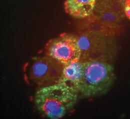 Once upon a time, in 2003, a French team discovered a giant virus infects amoeba. It was isolated from a cooling tower in the UK. They were so excited because it was so huge with a genome contains 900 protein-encoding genes (The words giant/ huge are totally hilarious. It’s not “Hulk”, it’s just a virus). It’s visible under the optical microscope. They named it Acanthamoeba polyphaga mimivirus (APMV). The prefix “mimi” is for mimicking microbe. Now, the same team “Raoult’s team” reported the isolation of another strain of those giant viruses but this time it was isolated from a cooling tower in Paris. They named it “mamavirus” because it was slightly larger than the previous giant virus (APMV), but it wasn’t alone. It was associated with its satellite, a small virus has 21 protein-encoding genes infects it, hijacks its viral factory making copies of itself, hindering the ability of the mamavirus to replicate/ make its own copies, so the number of the mamavirus drops in the infected amoebae. They named it Sputnik after the first man-made satellite. It’ll be the first isolated “Virophage”. How did I know about it? From the amazing blog of Dr. Ramy K. Aziz, “Microbes“. Once upon a time, in 2003, a French team discovered a giant virus infects amoeba. It was isolated from a cooling tower in the UK. They were so excited because it was so huge with a genome contains 900 protein-encoding genes (The words giant/ huge are totally hilarious. It’s not “Hulk”, it’s just a virus). It’s visible under the optical microscope. They named it Acanthamoeba polyphaga mimivirus (APMV). The prefix “mimi” is for mimicking microbe. Now, the same team “Raoult’s team” reported the isolation of another strain of those giant viruses but this time it was isolated from a cooling tower in Paris. They named it “mamavirus” because it was slightly larger than the previous giant virus (APMV), but it wasn’t alone. It was associated with its satellite, a small virus has 21 protein-encoding genes infects it, hijacks its viral factory making copies of itself, hindering the ability of the mamavirus to replicate/ make its own copies, so the number of the mamavirus drops in the infected amoebae. They named it Sputnik after the first man-made satellite. It’ll be the first isolated “Virophage”. How did I know about it? From the amazing blog of Dr. Ramy K. Aziz, “Microbes“.
The story won’t stop at this discovery. The discovery of the virophage will strongly suggest that “Viruses are alive” because they share something with other living domains of life, they can be infected, they can get sick, what makes all health-care providers totally thrilled because there’s something stronger than viruses which could be used to fight them, but “It’s too early to say we could use Sputnik as a weapon against big viruses or to modify them,” says co-author Bernard La Scola.
One more thing about Sputnik, 3 of its genes are closely related to APMV which suggests horizontal gene transfer between giant viruses caused by Sputnik. This is so “bacteriophagic”, reminds me with the whole insertion/ lysogenic mechanism between phages & bacteria. The isolated sequences from the ocean are closely related to the genome sequences of giant viruses & their satellite (Sputnik) . They infect plankton. “It suggests there are other representatives of this viral family out there in the environment,” Koonin says.
Image credits:
Giant mamavirus particles (red) and satellite viruses of mamavirus called Sputnik (green). http://www.nature.com/
Tags: APMV, horizontal gene transfer, mamavirus, sputnik, virophage
No Comments »
 U.S. Center for Disease Control and Prevention U.S. Center for Disease Control and Prevention
A new study reveals that people infected with bilharzia, or other parasitic worms, are more likely to become infected with HIV than normal persons. This was proven through an experiment where the infectious dose of an HIV-like virus necessary to infect rhesus macaques was found to be 17 times lower in animals with acute schistosomiasis than in controls. The animals co-infected with Schistosoma mansoni also showed higher memory cell concentrations of virus casuing a more rapid progression to AIDS.
These findings prove the assumption that persons living in highly endemic areas for parasitic worms have a higher risk of acquiring HIV/AIDS.
Previous studies by other research groups have demonstrated that the presence of schistosome infections increases viral replication in animal or human hosts with established immunodeficiency virus infections.
Both findings are surely to have profound public health implications for the under-developed areas of the world where both parasitic worms and HIV virus are endemic.
Tags: AIDS, bilharzia, HIV
10 Comments »
Nothing made the world highly concerned about the immune system, what are its components? How does it work?, better than the emergence of HIV in the 80s. It’s a disaster, but made us know more about the immune system. 
HIV’s target is CD4 receptors, which are present mostly on T-helper cells. It has glycoprotein 120 (Why do they call it 120 any way?! Is it the UV absorption again?! Or maybe it has 120 amino acids?!) It’s on its envelope. By recognition & binding to the CD4 receptors, it kills the T-helper which result in suppression of the whole cell-mediated immunity mechanism. It’s like cutting the snack’s head off. T-helper cells are responsible for giving signals (Interleukins) to other members of the IS so they can kill the viruses. By the whole suppression idea, the IS is turned off, the human body will be opened like your friend’s heart to you, to all possible invaders of m.o.
When we talked about HIV, the professor told us:” You wanna fight HIV, young docs full of enthusiasm, block its binding site, so it can’t bind to CD4 anymore.” I remembered the wise man’s words when I surveyed this article “Antibodies to the CD4-binding site of HIV-1 gp120 suppress gp120-specific CD4 T cell response while enhancing antibody response” about studying the effect of monoclonal anti-bodies against only the highly conserved part of the gp 120 (The binding site). We know that after exposure to HIV, the IS produces Ab against gp 120 to neutralize it, but the HIV tends to change the gp 120, so it can’t fit with the neutralizing Ab, moving on to more destruction. With those highly specific binders, I thought it’ll be the ultimate success.
Unfortunately, the research group made in vivo (in mice) & in vitro studies using the normal virus & another recombinant one with no CD4bs. They called it CD4bs+ Env & CD4bs- Env (Like with or without cheese). They found that the Anti-CD4bs mAb have high neutralizing activity, they raised the Ab titer (mainly IgG but not IgM). But they hinder the ability of the proteolytic enzymes/ the degrading mechanisms of phagocytes/ T-helper response to the envelope Ag/ the ability of Antigen presenting cells & MHC II to present the Ag. Let’s think about it…. They can only present the virus’s Ag, not the gp120/anti-CD4bs complexes. This is too long in writing, how about presenting? Just kidding, It’s about that the Ag is already covered, so it’s useless to be presented.
This is so awful, even the last approach to bind HIV didn’t work. What are the researchers gonna do? What’s the next move? We’ll find out soon.
Image credits:
Anatomy of AIDS virus: http://www.roshanpakistan.com/
Tags: HIV, immunity
2 Comments »
|

 Entries (RSS)