 For millions of years, our planet earth has stood still against the human race’s countless endeavors for destruction. Nevertheless, it has become apparent in recent years that earth’s defenses may be failing, and that through the uncontrollable use of fossil fuels, global warming strikes as earth’s faint cry for help. With humans being reluctant to listen, we are already witnessing the consequences. For millions of years, our planet earth has stood still against the human race’s countless endeavors for destruction. Nevertheless, it has become apparent in recent years that earth’s defenses may be failing, and that through the uncontrollable use of fossil fuels, global warming strikes as earth’s faint cry for help. With humans being reluctant to listen, we are already witnessing the consequences.
However, the answer to earth’s problems lies in plain sight to those who are wise enough to look. The scientists of East Anglia University and Ocean University China investigated marine microorganisms, a mysterious yet fascinating field that was often neglected in the past, their findings could be a promising step in controlling global warming.
A marine alphaproteobacterium, namely Labrenzia aggregata is a breakthrough discovery in the role of bacteria in the sulfur cycle and climate control.
It was previously thought that only eukaryotes contributed to the cycle, but Labrenzia aggregata was found to convert dimethyl sulfoniopropionate (DMSP) into dimethyl sulfide (DMS) through the methionine transamination pathway. DMSP is a nutrient for marine microorganisms and a precursor for DMS.
DMS plays a major role in climate control, it is oxidized in the atmosphere into sulfate aerosols which form cloud condensing nuclei that absorb UV radiation lowering atmospheric temperatures and counteracting the greenhouse effect, in addition, these clouds transfer sulfur from ocean to land contributing to the sulfur cycle.
Labrenzia aggregata is the first discovered heterotrophic bacterium that is able to produce DMSP through de novo production of methionine through its acquisition of the dsyB gene, which encodes a methyltransferase enzyme.
This provides further evidence that DMSP production is not restricted to phototrophs on the surface of the ocean, but extends through its entire depth.
Finally, the discovery that a single gene transfer between different strains allowed DMSP production is remarkable. Will this transfer enable us to recruit other heterotrophs in combating global warming? Could Alphaproteobacteria be our salvation or is it only a few Labrenzia strains? The possibilities are endless and the prospects are exciting! The world is eager to see what the ocean has yet to offer.
References:
https://www.nature.com/articles/nmicrobiol20179
https://www.sciencedaily.com/releases/2017/02/170213131452.htm
https://www.ncbi.nlm.nih.gov/pmc/articles/PMC134419/
Image source:
from: klimat.czn.uj.edu.pl (context)
Tags: alphaproteobacteria, DMSP
 Comments Off on Could an alphaproteobacterium be a silent global warming slayer?
 The term “superbug” is nothing new to the microbiology world but has only been under the spotlight for a few years, which consequently led to an increasing interest in antibiotic resistance by researchers. A superbug refers to a multidrug resistant bacterium which can therefore cause untreatable and fatal infections. This particular aspect has sparked global worry that our known antibiotics will eventually fail us. The term “superbug” is nothing new to the microbiology world but has only been under the spotlight for a few years, which consequently led to an increasing interest in antibiotic resistance by researchers. A superbug refers to a multidrug resistant bacterium which can therefore cause untreatable and fatal infections. This particular aspect has sparked global worry that our known antibiotics will eventually fail us.
With the interest in superbugs at its highest, scientists from Indiana University and Harvard University had their share in the investigation, using multi-colored dyes called fluorescent D-amino acids (FDAAs), aka rainbow dyes, which turned out to be just as cheerful to the researchers as actual rainbows. These dyes enabled them to visualize the detailed process of cell division, particularly the movement of the filaments FtsZ and FtsA (cytoskeletal polymers and prokaryotic homologs of the protein tubulin) that determine the site of cell division by driving peptidoglycan synthesizing enzymes to the correct sites. Cytokinesis starts with the formation of a Z-ring at the site of cell division, and both FtsZ and FtsA are required for this process.
When visualized, the filaments appeared to move in circular concentric rings, in a movement which was described as “treadmilling” in which the FtsZ filament loses a molecule at one end and gains a molecule at the other end, resulting in the circular motion. With the guidance of these rings, peptidoglycan was shown to begin forming a septum dividing the cell.
A more detailed aspect of the FtsZ and FtsA system is the lack of any means to convert chemical energy into mechanical force. However, the rearrangement is primarily dependent on FtsZ polymerization dynamics under the influence of conflicting regulation by FtsA, first, by promoting FtsZ assembly and second, by inhibiting FtsZ network organization. The result of this regulation is the formation of higher ordered structures by FtsZ as tubules, circles and sheets.
These findings might be a magnificent aid in combating superbugs by visualizing so accurately their division and offering a broader comprehension of their mechanisms.
Finally, if you ever find yourself anxious about superbugs, just remember: there always comes a RAINBOW after a rainy day.
References:
1 Comment »
 What could those three possibly have in common? Believe it or not, they all play the role of a lead action figure in kids video games. Hearing the latest edition of the german biotechnology news broadcast, I was surprised to learn that researchers at the Riedel-Kruse Lab in Stanford University have developed, what-they-call, Biotic Video Games, where the paramecium are controlled and maneuvered about via a joystick and managed to publish their findings in a research paper! What could those three possibly have in common? Believe it or not, they all play the role of a lead action figure in kids video games. Hearing the latest edition of the german biotechnology news broadcast, I was surprised to learn that researchers at the Riedel-Kruse Lab in Stanford University have developed, what-they-call, Biotic Video Games, where the paramecium are controlled and maneuvered about via a joystick and managed to publish their findings in a research paper!
The device is basically composed of a fluid compartment, where the paramecium move around and roam about freely. I am sure you are wondering, exactly how BIG (small) IS a paramecium. Well, it is so small, making it actually difficult to observe by the naked eye. But no worries! They can be seen quite clearly on your screen, while you’re playing, thanks to the provided microscope camera, which is connected to electrodes and supplies you with a live feed, being superimposed on the flash game board onto your screen. The joystick is capable of creating a weak electric field, which influences the direction of their movement, as you wish.
Eight different games have been developed and given quite funny names, as Ciliaball, Pac-man and Pond Pong. For instance, in one game version, the player needs to move about the paramecium to score a soccer goal. To help you easier imagine this, take a look at this 3-part video.
As Riedel-Kruse put it, these games serve two ultimate goals: First, to awaken the scientific interest in those young kids and teenagers, hopefully motivating them to someday pursue a career, heading off in that direction. And after all, scientists can collect and analyze information about those tiny organisms, whilst playing with them.
So please do take a break and enjoy some time away with your paramecium 🙂
Image Source: Stanford University Schools of Medicine and Engineering
 Riedel-Kruse IH, Chung AM, Dura B, Hamilton AL, & Lee BC (2011). Design, engineering and utility of biotic games. Lab on a chip, 11 (1), 14-22 PMID: 21085736 Riedel-Kruse IH, Chung AM, Dura B, Hamilton AL, & Lee BC (2011). Design, engineering and utility of biotic games. Lab on a chip, 11 (1), 14-22 PMID: 21085736
Tags: biotech game, biotic game, electric field, electrode, flash game, paramecium, super mario, super sonic, video game
No Comments »
Though risky, transplants are crucial treatment interventions for many patients.They can save lives of cancer patients, others with severely ill organs and recently there are trials to make them a mainstream treatment for autoimmune disease patients ( i’ve just read an article about that topic and i’d really love to write about it soon too).
But the major problem with transplants, other than the agonizing wait for the right donor on endless lists for sometimes many years, is that when things go wrong with transplants, the patient’s life becomes at mortal risk. Almost 40% of transplant patients will show rejection episodes within the first year after the operation. The detection of these immunological reactions are usually so late, and the only solution will be to flush the patient’s system with huge doses of immunosuppressive drugs that are toxic themselves and can have debilitating effects on cancer patients for instance. Also, to be able to detect rejection reactions, the doctors should take biopsies of the new organ, a process that can cause damage to the organ itself, let alone the stress and the already fragile patient condition. Transplant patients have to undergo exploratory biopsies monthly for one year after the operation!!!!!
 Will these risky, life-saving procedures be safer in the future? Early detection of these reactions was an interesting topic and a field of research for the cardiologist Hannah Valantine of Stanford University School of Medicine in Palo Alto, California. In 2009, she devised a new test that detects the immunological changes in a transplant patient in an episode of rejection. The test, called AlloMap, became the first of its kind to be approved by the FDA for use in the detection of heart transplant rejections. Yet, it failed to detect the rejections early in about half the patients.This, of course, didn’t satisfy Valantine.
Along with biophysicist Stephen Quake of Stanford, they came out with a much more sensitive test. The idea was that DNA from the new organ constitutes around 1% of the free DNA in blood of transplant patients. This DNA is foreign from the DNA of the patient and using her test, it can be very sensitively detected, despite the fact that it is circulating in minute amounts. To validate their test, they used it on stored plasma samples from transplant patients that later showed rejection signs. It was found that the amounts of the rejected organ’s DNA in such episodes are elevated soon after the surgery and constitutes around 3% of the free DNA in plasma, and of course, will be much elevated later, in the peak of the episode. They reported the results in The Proceedings of National Academy of Sciences.
The good thing about this test, besides its high sensitivity, is that it is much less invasive than a biopsy, and the biopsy will not be needed except for confirmation, in case the test is positive and the DNA % is higher than normal. Also, early detection will allow doctors to use much smaller doses of immunosuppressives to control the case and therefore, less side effects will be experienced. Valantine is a cardiologist, but believes the test can be used with other types of transplanted organs, other than hearts.
Source: ScienceMag
Tags: AlloMap, cardiology, FDA, immunosuppressives, organ rejection, transplants
No Comments »
 After the Human Genome Project was successfully completed in April 2003 and it was assured that humans are identical in the sequence of their genome by 99.9%, researchers are moving on to find more about the 0.1% left. Although our genome is made out of 3 billion bases (A’s, G’s, C’s and T’s), the 0.1% genetic difference is extremely significant. This is due to the fact that this small percentage holds the key to most of the consequences of such a variation between human beings (e.g susceptibility to diseases and also response to drugs). This introduced the term SNPs (single nucleotide polymorphism) which was found to be highly involved in variation of response to many drugs (like anti-cancers) and the list is increasing every day. After the Human Genome Project was successfully completed in April 2003 and it was assured that humans are identical in the sequence of their genome by 99.9%, researchers are moving on to find more about the 0.1% left. Although our genome is made out of 3 billion bases (A’s, G’s, C’s and T’s), the 0.1% genetic difference is extremely significant. This is due to the fact that this small percentage holds the key to most of the consequences of such a variation between human beings (e.g susceptibility to diseases and also response to drugs). This introduced the term SNPs (single nucleotide polymorphism) which was found to be highly involved in variation of response to many drugs (like anti-cancers) and the list is increasing every day.
To be more focused on the human variations represented by the 0.1 percent, the NIH led the international HapMap project. Their work was performed on four populations groups: the Yoruba people in Ibadan Nigeria,the Japanese in Tokyo, the Han Chinese from Beijing and Utah residents from western and northern Europe. The work began in October 2002 and successfully ended in October 2005. The work of three successive years helped the researchers invent a shortcut for studying SNPs. Scientists believe that there are about 10 million SNPs distributed among 3 billion base pairs which make up our genome, so scanning the whole genome of millions of people for such SNPs would be extremely expensive. After the HapMap project, researchers demonstrated that variants usually tend to cluster into neighborhoods (called haplotypes) and thus the number could be reduced to 300,000 SNPs only. This means that they could reduce the work load by about 30 folds.
The genome-wide association (GWA) studies aim to pinpoint the genetic differences, which cause a certain disease (or a biological trait) by comparing a group of people (who have the trait under research) to a control group (people who are free from this trait). Utilizing thousands of SNPs markers, we can identify regions (loci) which are statistically different between patient and control groups. Thus, we can identify the genetic difference between sick and control people, even though the difference was subtle. This means that the combination of slightly altered genes plus environmental factors could be well studied. The conventional ways, usually used to study genetic differences, are mainly based on selecting the candidate gene based on knowing or suspecting the mechanism of the disease. GWA helps scanning of the whole genome in a comprehensive unbiased manner. It will let us get the whole picture about other, non expected, contributing genes. In this way, GWA studies will help us study the multi-factorial diseases (like cancer and diabetes) in a more rationalised way.
Another challenge has come up: What about the genetic variations due to geographic ancestry? It is also a significant contributing factor to variation among humans and all the efforts are directed towards making a somewhat universal map of human genome to help develop individualized drugs. A group of scientists led by David Reich, an assistant professor at Harvard Medical School, described a quantitative method that can correct such errors due to geographical ancestry known collectively as “population stratification”. It will help if the disease groups, sharing the same trait, have differences in their geographic ancestry.
Tags: Genome wide association studies, Genome-wide association studiesX, GWA, GWAS, Hap-Map project, HGP, human variome project, SNPs, variome
No Comments »
Infections in the teeth implants are dreaded, running a high risk of potentially affecting the jaw bones. It is actually not that uncommon. The overall number of performed teeth implantations in the US and Europe has doubled over the last decade, yet studies show that 10 percent of the implants are associated with problems, usually in the first year directly after the operation. To prevent any further deterioration, the implants sometimes have to be removed. teeth implantations in the US and Europe has doubled over the last decade, yet studies show that 10 percent of the implants are associated with problems, usually in the first year directly after the operation. To prevent any further deterioration, the implants sometimes have to be removed.
New methods are now being developed by researchers in the University of Zurich to keep inflammation-causing bacteria at bay. In their PLoS ONE article, they present their materials and methods, which have successfully eliminated 99% of the microbes after a 15-minute electrical treatment.
Conventional treatment methods of this sort of inflammation depend on the utilization of topical antibiotics, which is surely a burden for the patient. The aim of the study was to develop a non-invasive approach to efficiently fight off the bacteria, or, as the researchers phrase it in their paper, to develop “an in-situ decontamination of the dental implants”.
The whole idea is based on the process of water treatment, where sterile water is produced through electrolysis. In order to simulate the conditions in the jaw, an Escherichia coli bacterial film was coated onto the titanium implants, which were impregnated in a gelatinous preparation. In the experimental design, one implant functioned as the cathode and the other as the anode. The implants were subjected to a 15-minute-long electrical treatment, of an intensity ranging between 0 and 10 Milliampere. This artificially generated electrical field caused the hydroxyl ions of the water molecules to migrate to the cathode, and thus raising the pH. A color change of the indicators, used in the gelatin, prove that an alkaline environment predominates at the cathode. On the other hand, the pH value drops at the anode, forming an acidic milieu.
The numerous experimental models with various electrical intensities show that in cases, where an acidic ambience was produced around the implants, 99% of the bacteria died off after a 15-minute treatment. Therefore, the patient implants in the future will take up the role of the anode. A clip at the lip will be used as a cathode.
What at first glance might seem as a torture mechanism is in reality completely harmless. The minute amount of Milliampere, which is sufficient to conquer the bacteria, is hardly even perceived by the patient and would tops cause a mild muscle twitching.
Tags: electrolysis, escherichia coli, PLoS ONE, teeth implant, Universität Zurich, zurich university
3 Comments »
If you have ever been one of the unlucky ones waiting for a cancer diagnosis biopsy, or having a friend or a relative undergoing the process, then you must know how the wait is nerve wrecking. The standard procedure is using a biopsy needle to extract some cells of the tissue suspected for cancer, and then waiting for about a week, until the results come out. To make matters worse, results can sometimes be inconclusive or 100% correct.
 Simple smartphone applications might be able to rapidly diagnose cancer in the future Fortunately, a group of scientists at Massachusetts General Hospital (MGH) in Boston, were able to develop a new technology, that is much more rapid and almost 100% accurate in the diagnosis process. They developed a small NMR device (detects compounds by the mode of oscillation of their nuclei in a magnetic field) the size of about a coffee cup, and they were also able to synthesize magnetic Nanoparticles, which stick to certain tumor specific proteins. So now all I have to do, is head to the clinic, have the needle biopsy performed and the cells taken. Then they are mixed with the magnetic Nanoparticles and the results are taken from the small NMR device and read using a simple smartphone application.
This technique was used in the first trial on 50 patients, taking less than an hour to diagnose each. Also, as the device can detect 9 tumor associated proteins, combining the results for 4 of these gave accurate results in 48 out of the 50 patients. In another trial, the accuracy was 100% in the 20 patients tested. The conventional tests’ average accuracy is 74-84 %.
This new technology will also cut down on the cost of repeat biopsies, which can be very expensive, and scientists hope it will have many other applications as well, like patient cancer follow-up, through quantitative analysis of the tumor associated proteins. Maybe the biopsy will not be needed in the future and a simple blood test will also do….
Source: Science/AAAS
Tags: cancer, cancer diagnosis, magnetic nanoparticles, NMR, smart phones, tumor associated proteins
2 Comments »
Metagenomics is a culture independent approach that has contributed extensively to the study and understanding previously unidentified microbial communities. Seeking a further understanding of employing metagenomics in the study of the Red Sea microbial communities, we are pleased to interview Dr. Rania Siam, an Associate Professor in the Biology Department, the Director of the Biotechnology Graduate Program at the American University in Cairo (AUC) and an Investigator in the Red Sea Marine Metagenomics Project that is currently running at the AUC in collaboration with King Abdullah University of Science and Technology (KAUST), Woods Hole Oceanographic Institution and Virginia Bioinformatics Institute at Virginia Tech. Dr. Siam holds a Ph.D. in Microbiology and Immunology from McGill University. In addition, she held several post-doctoral positions at McGill Oncology Group, Royal Victoria Hospital, The Salk Institute for Biological Studies and The Scripps Research Institute. Since 2008, Dr. Hamza El Dorry (PI) and Dr. Siam (Co-PI) have been leading the Red Sea Metagenomics research team. The team is exploring novel bacterial communities in the Red Sea through actively participating in Red Sea expeditions for sampling and performing extensive molecular biology, genomics and computational analysis of the data.

1- Dr. Siam, thank you very much for accepting our request. Would you please explain for us the driving motives behind doing metagenomics research in the Red Sea?
The Red Sea is a unique environment in the region that remains to be explored. Thus, working on the Red Sea gives us the opportunity to perform essential research for the region. Furthermore, our main interest are the Red Sea brine pools that are unique environments in terms of high temperature, high salinity, high metal contents and low oxygen. Microbial communities living in these environments are known as extremophiles. The survival of extremophiles in such drastic conditions indicates their possession of genes with novel properties that underlie these unique survival characteristics. Thus, we are highly motivated to explore these novel microbial communities and their unique properties. In addition, we are interested in extracting biotechnological products from the Red Sea that can be beneficial as antimicrobials and anticancer agents.
2- Would you please outline for us the objectives of the project and the main activities inside and outside the laboratory?
In our project, one of our main aims is establishing a Red Sea marine genomic database to be accessed by scientists’ worldwide. Additionally, we are screening this database for biotechnological pharmaceutical products as enzymes and anticancer agents. There are three main activities in the project: sampling, molecular biology/genomics work and computational analysis of the data. Concerning sampling, it is a challenging process that requires rigorous planning where samples should be subjected to proper processing and storage till arrival to the labs. In labs, samples are subjected to different procedures starting from DNA extraction followed by Whole Genome Sequencing (WGS) to identify unknown genes or unique ones and help us understand novel microbial communities. This requires rigorous computational analysis to make sense of our data. Furthermore, we construct fosmid libraries for isolation and purification of genes of biotechnological interest as lipases and cellulases. In addition, we carry out 16s rRNA phylogenetic analysis on the microbial communities present in the samples.
3- Since the idea is novel, we would like to know about the nature of samples, the parameters and the challenges imposed during the process of sampling.
Basically, the sampling process requires well-equipped research vessels as the Woods Hole Oceanographic Institute ‘Oceanus’ and The Hellenic Center for Marine Research (HCMR) ‘Aegeo’. In addition, it is essential to have a team of physical oceanographics for adequate sampling.

http://www1.aucegypt.edu/publications/auctoday/AUCTodayFall09/Cure.htm
Image Source: AUC Today
We started with two different brine pools: Atlantis II Deep and Discovery Deep. As brine pools, these two regions are characterized by the presence of extremely harsh conditions as I mentioned before. In the 2008 and the 2010 KAUST Expedition to the Red Sea, we collected two forms of samples: Large volume water and sediments. In both cases, we face challenges during sample collection. Collecting large volume water samples can take up to 4 hours. In case of bad weather, it is nearly impossible to collect samples. Regarding the sediments, the heavy weight of the sediment core is the main challenge since it may drag people to the water during the sampling process.
The water samples are collected using CTDs (an acronym for Conductivity, Temperature and Depth), it is formed of 10 liter bottles that collect samples and measure the conductivity, temperature and depth, in addition to other parameters. CTDs are capable of measuring these physical properties from each meter of water. Accordingly, they are able to retrieve 2200 readings for each parameter at 2200 m depth. This is very beneficial as it allows us to correlate the physical and chemical parameters with the nature of microbial communities obtained from each sample.
4- What are the reasons behind the choice of Atlantis II Deep and Discovery Deep brine pools for study?
These sites are unique. Atlantis II Deep is 2200 meters below the water surface. It is characterized by having high temperature (68 °C), high heavy metal content, high salinity and little oxygen. Thus, these extremely drastic conditions are motivating us to explore the extremophilic microbial communities in this pool. Discovery Deep is adjacent to Atlantis II but the conditions are less harsh and varies in its heavy metal content. This encourages us to undergo comparative genomic analysis between the two regions.
5- Did the relatively new metagenomic approach of sequencing multiple genomes, combined with, the novelty of employing this approach in the study of microbial milieu in the Red Sea imply some unprecedented practical challenges in retrieving entire and authentic DNA sequences?
Yes, many challenges are present. For example, in case of sediments, many cells die. This in turn can make the process of DNA extraction more difficult. However, we managed to cope with this problem by quickly extracting the DNA from the sediments following its arrival to the labs and avoid freezing and thawing. Another problem with sediments is the presence of impurities that are co-extracted with DNA and interfere with the analysis. Concerning water samples, the main challenge is that the amount of DNA in the samples is very minute. This was dealt with by filtering large volumes of water up to 500 liters per sample.
6- Is recognizing and validating sets of data that are pointing out to specific patterns of microbial diversity or novel genes considered to be challenging?
Actually, we have retrieved enormous amount of data and a large percentage of the data has no match to sequences present in the other genomic databases. This has inspired us to think of new approaches for data analysis to identify the role of these novel sequences and seek collaborations with computational biologists.
7- Finally, do you think there are other environments in Egypt with unique properties which make it promising for employing metagenomics to discover novel genes and bacterial strains?
Yes, actually Egypt is very rich in environments with unique properties. For example, we have the deserts, Siwa‘s hot springs and the Nile. Many unique environments are yet to be explored and lots of research needs to be performed. Our natural resources are limitless.
Tags: 16s rRNA phylogenetic analysis, Aegeo, Atlantis II, AUC, Brine pools, Computational analysis, Computational biologists, CTDs, Culture-independent, Discovery Deep, DNA extraction, Drastic conditions, Expeditions, Extremophiles, Fosmid libraries, genomics, KAUST, Metagenomics, microbial communities, Molecular biology, Oceanus, Red Sea, Virginia Bioinformatics Institute, Virginia Tech., WGS, Woods Hole Oceanographic Institution
2 Comments »
 A 70-year-old man can enjoy the blessing of fatherhood to perfectly healthy children. That is practically a miracle quality-wise because the male gametes are being produced in huge amounts, 1000 per second to be exact. That amounts to about 30 billion a year. Each and every one of those sperms could potentially contribute by half to the formation of a human being. So how could this rapid production preserve and maintain the critical quality required? How could errors during the sperm production be avoided? Researchers have recently gained an insight into the molecular mechanism as released in the “Proceedings of the National Academy of Sciences”. A 70-year-old man can enjoy the blessing of fatherhood to perfectly healthy children. That is practically a miracle quality-wise because the male gametes are being produced in huge amounts, 1000 per second to be exact. That amounts to about 30 billion a year. Each and every one of those sperms could potentially contribute by half to the formation of a human being. So how could this rapid production preserve and maintain the critical quality required? How could errors during the sperm production be avoided? Researchers have recently gained an insight into the molecular mechanism as released in the “Proceedings of the National Academy of Sciences”.
During the sperm production, there is an automatic quality control process. This control mechanism is strengthened by a specific genetic addition, present in both humans and great apes. The triggering factor is comprised of parts of an endogenous retrovirus, incorporated in our genome. 15 million years ago, this viral DNA was presumably incorporated in the genetic makeup of one of our ancestors. A lucky coincidence? Perhaps, but the researchers claim it catched on during the process of evolution. The site of insertion of this viral DNA is close to a gene, responsible for the production of a crucial control factor.
The control factor, termed p63, drives faulty cells straight to their apoptosis. It imposes a strict quality control of the genome because even in cases of a slight damage to the DNA, the cells ultimately die. As a result, the passing on of a flawed genome to the next generation is prevented. Some cells definitely fall as victims in the process. In addition, this mechanism could protect against certain types of cancer as testicular carcinoma. They refer that, p63 represents a barrier to the formation of tumors in normal healthy tissues. In cases of testicular cancer, the administration of drugs, that restore the function of p63, could prove potentially useful in the near future.
Source: Pharmazeutische Zeitung and original PNAS paper (thanks to Mariam and Steve Moss for sharing)
Tags: evolution, p63, PNAS, quality control, retrovirus, sperm, testicular carcinoma, tumor
3 Comments »
 A simple online search for anti-microbial clothing and you are bound to come across an assortment of brand names, even Adidas, promoting their products, mostly sportswear. But are they really safe? To answer this question, researchers at the Hohenstein Institute tested the safety of such anti-bacterial textiles. The result: Even in cases of volunteers, who wore those clothing articles for an extended period of time, their skin flora was not affected. A simple online search for anti-microbial clothing and you are bound to come across an assortment of brand names, even Adidas, promoting their products, mostly sportswear. But are they really safe? To answer this question, researchers at the Hohenstein Institute tested the safety of such anti-bacterial textiles. The result: Even in cases of volunteers, who wore those clothing articles for an extended period of time, their skin flora was not affected.
Over the last years, anti-microbial textiles have gained popularity. It started off in the medical sector in an approach to cut down on cross-infection between patients and to ensure the safety of the hospital staff. Recently, the industry has managed to find potential customers, those who are interested in sport and outdoor activities, looking for ways to control bad odours. The majority of these products, found on the market, are manufactured using anti-bacterial silver strands, which help in “eliminating 99.9% of odour-causing bacteria” as one ad claims. Furthermore, one firm releases products to be used in cases of neurodermatitis. On their homepage, it is stated that, “our silver textiles provide a realistic alleviation of this agonizing skin disease” and are backed up by statements of health experts.
Despite the previous positive experiences concerning the implementation of silver, like in the purification of water, anti-bacterial clothing have raised a controversial discussion in the media. This encouraged researchers to run a 6-week-long experiment, in which 60 healthy volunteers participated. For the study, special T-shirts were manufactured, which conveyed antimicrobial activity on one side, yet a non-antibacterial placebo effect on the other. For confirmation that this model actually did exert an antibacterial effect, the T-shirts were tested in the laboratory using Staphylococcus aureus und Klebsiella pneumonia. The volunteers were instructed to wear the T-shirts for at least 8 hours each day during the course of 4 weeks. The researchers analyzed on a weekly basis several parameters concerning the skin flora. In all the participating volunteers, the skin flora, both before and after, were within the normal range. There were no threats of a skin pathogen at any time. Therefore, these clothes have been characterized as being harmless and it seems like, their market will continue to expand as more people appreciate the benefits of leading an active life style.
Tags: adidas, antibacterial, antimicrobial, bad odour, hohenstein, silver
No Comments »
|
 For millions of years, our planet earth has stood still against the human race’s countless endeavors for destruction. Nevertheless, it has become apparent in recent years that earth’s defenses may be failing, and that through the uncontrollable use of fossil fuels, global warming strikes as earth’s faint cry for help. With humans being reluctant to listen, we are already witnessing the consequences.


 Entries (RSS)
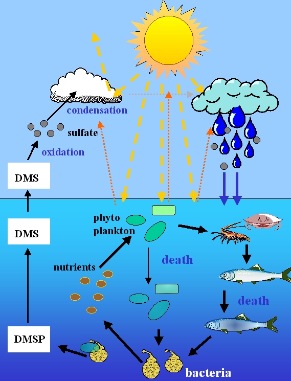